Introduction
Total blood volume in an adult: 5-6 L or 7-8% of the total body weight
Liquid Portion: 55% = 90% water, 10% proteins (albumin, fibrinogen, globulin)
- Blood Plasma
- liquid portion if coagulation is prevented
- formed elements can be separated
- unclotted blood contains fibrinogen
- Serum
- liquid portion expressed from the clot devoid of fibrinogen
- lacks coagulation factors I, V, and VIII
Solid Portion: 45%
- Made up of formed elements known as hemocytes (RBC, WBC, and Platelets)
Formation of Blood
- Plasma is derived from the intestines and organs of the body
- Formed elements are derived from the bone marrow of the bones.
Physical Characteristics
- It is fluid state in vivo
- Is red in color due to hemoglobin
- It is slightly alkaline (pH 7.4)
- Has an average spGr of 1.055 (1.045 – 1.065)
- It is thick and viscous (3.5 – 4.5x thicker than water)
- It contains approximately 20gms solid per 100ml of blood
- It makes up 75 – 85ml blood per kg body weight
Functions of Blood
- It carries O2 from the lung to the tissues and CO2 from the body tissues to the lungs.
- It conveys raw materials of the protoplasm to countless millions of cells as factories to manufacture life itself.
- It carries waste products to organs of elimination.
- It transfers hormones from their organs of production to the organs of consumption.
- It aids in regulating the H2O content, temp, and alkalinity of the tissues.
- It helps in protecting the body to battle infection and disease.
- It furnishes factors of coagulation to mend an abrasion, laceration and incision.
Blood Volume
- The total amount of blood in the circulation.
- Total blood volume (TBV) is the sum of the red cell volume (RCV) and plasma volume (PV) (TBV = RCV + PV)
Two Principal Factors for Maintaining a Normal Blood Volume
- Capillary Fluid Shift Mechanism
- If there is an increase of fluid in the blood, it automatically leaks into the tissue spaces
- If blood volume decreases, the fluid is absorbed from the interstitial spaces.
- Kidney Regulation Mechanism
- Excretion of excess fluids and the reabsorption in the tubules.
Blood Volume Determination
- To evaluate blood and body fluid losses during major surgical procedures and following trauma.
- To serve as a guide in pre and post operative transfusion therapy.
- To evaluate gastrointestinal and uterine bleeding.
- To aid in the diagnosis of hypovolemic shock1.
- To aid in the diagnosis of polycythemia vera2.
Causes of Hypovolemic States:
- Loss of WBC, RBC, plasma, body fluid or H2O
- Surgical shocks, nephrotic syndrome and peritoneal analysis
Causes of Hypervolemic states:
- During excessive fluid intake
- During intravenous injection of fluid like dextrose
- Blood transfusion
- During pregnancy
General Methods of Obtaining Blood
- Skin puncture
- Venipuncture
- Arterial puncture
Whole Blood

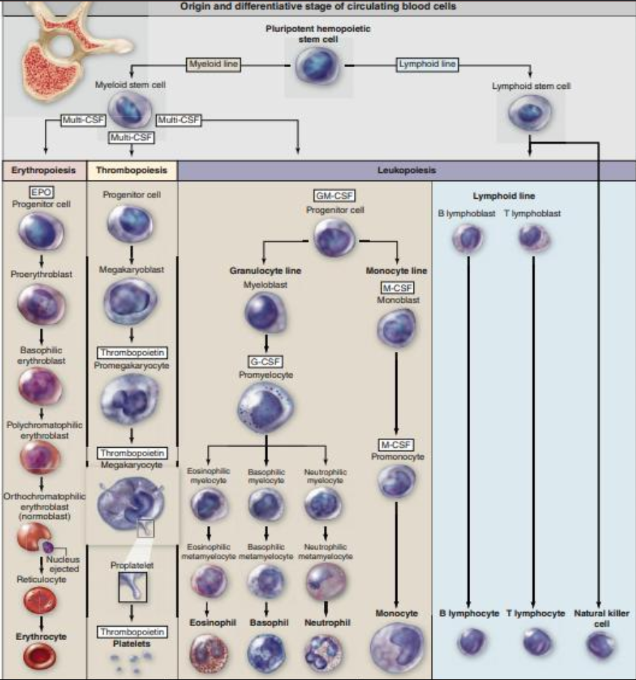
Hemopoiesis
- In the embryo blood cells arise in the yolk sac.
- In the 2nd trimester, it occurs primarily in the developing liver and spleen.
- In the 3rd trimester, marrow of specific bones becomes the major hemopoietic organ.
- After birth, hemopoiesis continues almost exclusively in the red marrow of different bones.
- The origin and maturation of cells are termed, erythropoiesis, granulopoiesis, monocytopoiesis, and thrombocytopoiesis.
- Lymphopoiesis or lymphocyte development occurs in the marrow and in the lymphoid organs.
- Pluripotential hemopoietic stem cell
- cell that can produces all blood cell types.
- produce: pluripotential myeloid stem cells and pluripotential lymphoid stem cells.
- Myeloid stem cells
- develop in red bone marrow
- erythrocytes, eosinophils, neutrophils, basophils, monocytes, and megakaryocytes
- Lymphoid stem cells
- develop in red bone marrow
- some lymphoid cells remain in the bone marrow to become B lymphocytes.
- other leave the bone marrow and migrate via the bloodstream to the lymph nodes and spleen.
- other undifferentiated lymphoid cells migrate to the thymus gland, where they differentiate into T lymphocytes.
- The progenitor cells for blood cells are often called colony forming units (CFUs).
- Four major types of progenitor cells/CFUs:
- Erythroid lineage - erythrocytes.
- Thrombocytic lineage - megakaryocytes for platelet formation.
- Granulocyte-monocyte lineage - granulocytes and monocytes.
- Lymphoid lineage - B lymphocytes, T lymphocytes, and natural killer (NK) cells.
- Colony stimulating factors (CSF) or cytokines
- hemopoietic growth factors
- glycoproteins that stimulate proliferation of progenitor and precursor cells and promote cell differentiation and maturation within specific lineages.
| Cytokine | Major Activities and Target Cells |
|---|---|
| Erythropoietin (EPO) | Mitogen for all erythroid progenitor and precursor cells, also promoting their differentiation |
| Thrombopoietin (TPO) | Mitogen for megakaryoblasts and their progenitor cells |
| Granulocyte-macrophage colony-stimulating factor (GM-CSF) | Mitogen for all myeloid progenitor cells |
| Granulocyte colony-stimulating factor (G-CSF or filgrastim) | Mitogen for neutrophil precursor cells |
| Monocyte colony-stimulating factor (M-CSF) | Mitogen for monocyte precursor cells |
Erythropoiesis
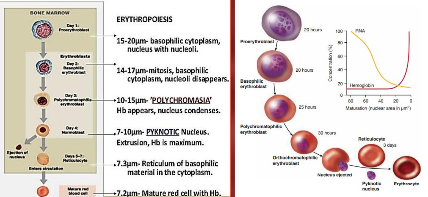
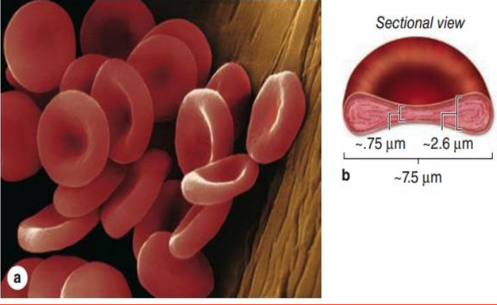
- Nonnucleated biconcave disks that are surrounded by a membrane and filled with hemoglobin and some enzymes.
- Oxyhemoglobin - is responsible for the bright red color of arterial blood.
- Carbaminohemoglobin - gives venous blood its bluish color.
- They are uniform in size and measure approximately 7.5 μm in diameter.
- RBCs are the only blood cells whose function does not require them to leave the vasculature.
- The life span is approximately 120 days.
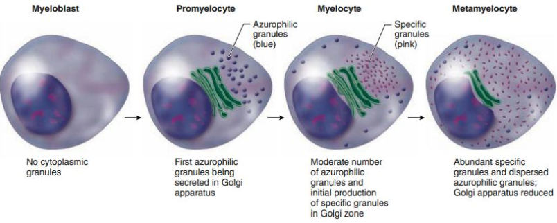
Granulopoiesis
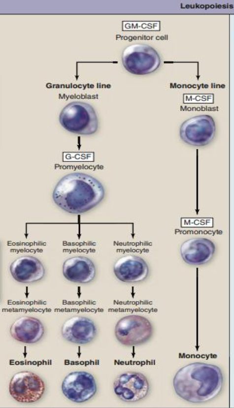
Synthesis of proteins for the azurophilic granules and specific granules.
- The myeloblast is the most immature recognizable cell in the myeloid series.
- The promyelocyte is characterized by basophilic cytoplasm and azurophilic granules containing lysosomal enzymes and myeloperoxidase.
- The first visible sign of this differentiation appears in the myelocyte stage.
- Specific granules gradually increase in number and eventually occupy most of the cytoplasm at the metamyelocyte stage.
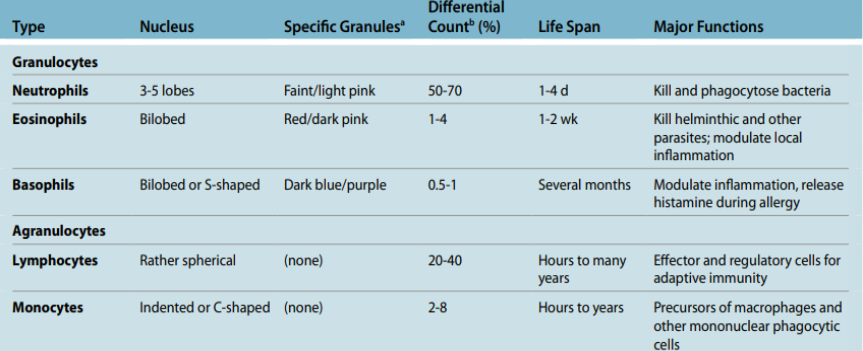
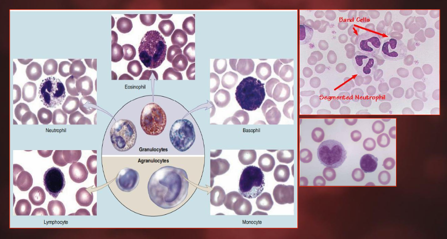
Neutrophil
- Mature neutrophils constitute 50%-70% of circulating leukocytes.
- Neutrophils are 12-15 μm in diameter in blood smears, with nuclei having two to five lobes linked by thin nuclear extensions.
- Neutrophils are inactive and spherical while circulating but become amoeboid and highly active during diapedesis3.
- Usually the first leukocytes to arrive at sites of infection where they actively pursue bacterial cells using chemotaxis and remove the invaders or their debris by phagocytosis.
- Azurophilic primary granules: Myeloperoxidase (MPO), Lysozyme & Defensins.
- Specific secondary granules: are smaller and less dense, stain faintly pink, and have diverse functions.
Stages
- Myeloblast - Large cell with a large nucleus and which demonstrates basophilic staining.
- Promyelocyte - During this stage primary (azurophilic) granules are formed.
- Neutrophilic myelocyte - The developing neutrophil can now be differentiated from basophils and eosinophils as neutrophil specific granules are now being formed.
- Neutrophilic metamyelocyte - At this stage mitosis can no longer occur. The nucleus elongates, becomes heterochromatic and has a kidney like shape.
- Band cell - Nucleus elongates further and represents a horse shoe. Nucleus starts to segment.
- Neutrophil - Mature neutrophil is formed and the nucleus is segmented and has 3 to 5 lobes. This lobular structure of the nucleus gives rise to the name polymorphonuclear neutrophil.
Eosinophil
- Constituting only 1%-4% of leukocytes.
- The nucleus is typically bilobed, but a small third lobe may be present.
- The main identifying characteristic is the abundance of large, acidophilic specific granules typically staining pink or red.
- Major basic proteins (MBP) along with eosinophilic peroxidase, other enzymes and toxins, act to kill parasitic worms or helminths.
- Also modulate inflammatory responses by releasing chemokines, cytokines, and lipid mediators, with an important role in the inflammatory response triggered by allergies.
Stages
- Myeloblast (1) & Promyelocyte (2) - These stages are common to all granulocytes and no distinction can be made between different cell lines.
- Eosinophilic myelocyte (3) & metamyelocyte (4) - Specific granules start to appear in the myelocyte stage and once the cell has reached the metamyelocyte stage it cannot undergo further mitosis.
- Eosinophil (6) - Mature cell has a bilobed nucleus. There are species specific variations in granule size once stained.
Basophil
- Make up less than 1% of circulating leukocytes.
- The nucleus is divided into two irregular lobes, but the large specific granules overlying the nucleus usually obscure its shape.
- The granules in basophils are not as numerous as in eosinophils, however they are more variable in size, less densely packed, and stain dark blue or brown.
- Basophilic specific granules also contain much histamine and various other mediators of inflammation.
Stages
- Myeloblast (1) & Promyelocyte (2) - These stages are common to all granulocytes and no distinction can be made between different cell lines.
- Basophilic myelocyte (3) & metamyelocyte (4) - Specific granules start to appear in the myelocyte stage, and as the cell develops into the metamyelocyte stage, mitosis ceases.
- Basophil (5) - Final nuclear shape is masked by the high density of cytoplasmic granules.
Monocyte
- Monocytes are agranulocytes that are precursor cells of macrophages, osteoclasts, microglia, and other cells of the mononuclear phagocyte system in connective tissue.
- All monocyte-derived cells are antigen-presenting cells and have important roles in immune defense of tissues.
- The monocyte nucleus is large and usually distinctly indented or C-shaped.
- The cytoplasm of the monocyte is basophilic and contains many small lysosomal azurophilic granules.
- These granules are distributed through the cytoplasm, giving it a bluish-gray color in stained smears.
Monocytopoiesis
- Monoblast - This is the first stage after cell has differentiated into the CFU-M.
- Promonocyte - Cell has a large nucleus and basophilic cytoplasm and consists of two populations: One rapidly dividing and the other slowly dividing, which acts as a reservoir.
- Monocyte - Monocytes are incapable of mitosis and enter the circulation. They have a large kidney shaped nucleus with a slightly basophilic cytoplasm, which is often vacuolated.
- Macrophage - Once the monocyte has entered tissue it differentiates into a macrophage.
- Dendritic cells - These develop from the monoblast under the stimulation of GM-CSF and IL-4 into an immature dendritic cell. This then develops into the mature dendritic cell under stimulation of TNF-α4.
Lymphocyte
- The most numerous type of agranulocyte in normal blood smears with a spherical nuclei.
- They are typically the smallest leukocytes.
- Major classes include: B lymphocytes, T lymphocytes, and natural killer (NK) cells.
- Have diverse roles in immune defenses against invading microorganisms and certain parasites or abnormal cells.
- Circulating lymphocytes have a wider range of sizes than most leukocytes.
- Small lymphocytes are characterized by spherical nuclei with highly condensed chromatin and only a thin surrounding rim of scant cytoplasm.
- Larger lymphocytes have larger, slightly indented nuclei and more cytoplasm that is slightly basophilic, with a few azurophilic granules, mitochondria, free polysomes, and other organelles.
Lymphopoiesis
T Cells
- T cells are formed in bone marrow then migrate to the cortex of the thymus to undergo maturation in an antigen-free environment for about one week where a mere 2-4% of the T cells succeed.
- Upon maturity, there are several forms of thymocytes including:
- T-helper (needed for activation of other cells such as B cells and macrophages),
- T-cytotoxic (which kill virally infected cells),
- T-memory (T cells that remember antigens previously encountered), and
- T-suppressor cells (which moderate the immune response of other leukocytes; also called T-regulatory cells (Tregs))
B Cells
- B cells are formed and mature in bone marrow (and spleen).
- Once in a secondary lymphoid organ, the B cell can be introduced to an antigen that it is able to recognize.
- Through this antigen recognition and other cell interactions the B cell becomes activated and then divides and differentiates to become a plasma cell.
- The plasma cell, a B cell end product, is a very active antibody-secreting cell that helps protect the body by attacking and binding to antigen.
- B lymphocytes are identified by the presence of soluble immunoglobulin G (IgG).
Summary
| WBC | Percentage | Actions |
|---|---|---|
| NEUTROPHILS | 50-70% | Neutrophilia - bacterial infection. Neutropenia - viral infections. |
| EOSINOPHILS | 1-3% | Eosinophilia - allergy or parasitic infection. |
| BASOPHIL | 0-2% | Basophilia - hematologic disease. |
| LYMPHOCYTES | 18-42% | Lymphocytosis - viral infections. Lymphocytopenia - drug therapy or immunodeficiency. |
| MONOCYTES | 2-11% | Monocytosis - infections, collagen-vascular diseases, or in acute and chronic leukemias. |
Thrombocyte

- Blood platelets are very small, non-nucleated, membrane-bound cell fragments only 2-4 μm in diameter.
- Normal platelet counts range from 150,000 to 400,000/μL (mm3 ) of blood.
- Circulating platelets have a life span of about 10 days.
- In stained blood smears, platelets often appear in clumps. Thrombocytopoiesis
- Megakaryoblast - Cell is slightly basophilic, around 30μm and has a round nucleus that is non-lobed.
- Promegakaryocyte - Cell is around 45μm with a larger cytoplasm and nucleus.
- Megakaryocyte - Cell is around 50-70μm and responds to thrombopoietin (TPO) and undergoes endomitosis. The size of the cell’s cytoplasm and nucleus increases with the increase in the cell’s chromosome number.
- Thrombocytes
Role of Platelets
- Primary aggregation: a platelet plug is formed as a first step to stop bleeding.
- Secondary aggregation: Platelets in the plug release a specific adhesive and increase the size of the platelet plug.
- Blood coagulation: more platelets to form a blood clot, or thrombus.
- Clot retraction: The clot that initially bulges into the blood vessel lumen contracts.
- Clot removal: Protected by the clot, the endothelium and surrounding tunic are restored by new tissue, and the clot is then removed, mainly dissolved by the proteolytic enzyme plasmin.
Picture of Cells
Footnotes
-
Hypovolemic shock is a medical emergency in which you’ve lost so much blood or fluid, your body can’t send enough of it to all of your organs. (Source) ↩
-
In oncology, polycythemia vera is an uncommon myeloproliferative neoplasm in which the bone marrow makes too many red blood cells. It is a type of chronic leukemia/blood cancer. (Source) ↩
-
Diapedesis is a rapid process in which the leukocyte extends itself by a pseudopod across the endothelial border. (Source) ↩
-
Tumor Necrosis Factor alpha (TNF-α) is a pro-inflammatory cytokine that has various effects in the response to injury and infection, angiogenesis, apoptosis, and other physiological processes. (Source) ↩